Read about Positron Emission Tomography (PET)
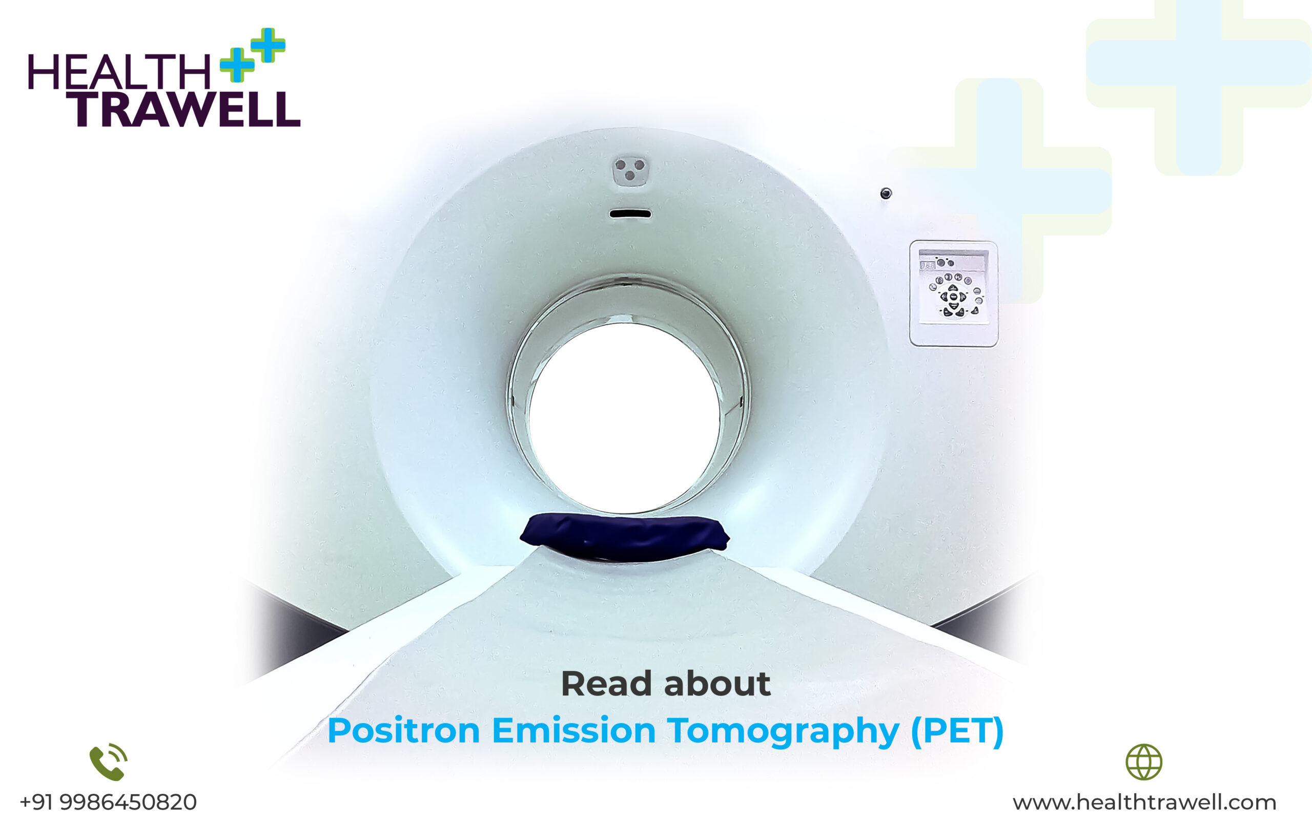
Positron Emission Tomography (PET)
Positron Emission Tomography (PET) is a medical imaging technique that produces detailed images of the internal structures and functions of the body. PET is a non-invasive diagnostic tool that can detect changes in cell activity and metabolism, and it is used to diagnose and monitor various diseases, including cancer, neurological disorders, and heart disease.
How Does PET Work?
Positron Emission Tomography (PET) works by using a radioactive substance called a tracer that is injected into the patient’s body. The tracer emits positrons, which are positively charged particles that interact with electrons in the body’s tissues. When a positron collides with an electron, the two particles annihilate each other, releasing gamma rays in the process. The gamma rays are detected by a scanner that is positioned around the patient’s body. The scanner produces detailed images of the distribution of the tracer in the body, which can be used to identify areas of abnormal cell activity.
Why Is PET Performed?
Positron Emission Tomography (PET) is used to diagnose and monitor various diseases, including cancer, neurological disorders, and heart disease. PET can help identify the location and extent of tumours, as well as determine whether cancer has spread to other parts of the body. PET can also be used to assess the effectiveness of cancer treatment and monitor for recurrence. PET can also be used to diagnose and monitor neurological disorders, such as Alzheimer’s disease, Parkinson’s disease, and epilepsy. PET can help identify areas of the brain that are affected by these conditions, which can help with treatment planning and monitoring. PET can also be used to assess heart function and blood flow. PET can help identify areas of the heart that are not receiving enough blood flow, which can be an indicator of heart disease. PET can also help assess the effectiveness of heart treatments, such as angioplasty or bypass surgery.
How Is PET Performed?
Positron Emission Tomography (PET) is performed in a specialized imaging facility that has the necessary equipment and trained personnel. Before the PET scan, the patient is injected with a small amount of a radioactive tracer. The tracer is usually administered intravenously, although it can also be inhaled or swallowed, depending on the type of test being performed. The patient then rests for a period of time, typically between 30 and 60 minutes, to allow the tracer to circulate throughout the body. The patient is then positioned on a scanning bed, and the scanner is positioned around the body. The scan itself usually takes between 20 and 45 minutes, depending on the area being imaged. During the scan, the patient must remain still to avoid blurring of the images. The patient may be asked to hold their breath for short periods during the scan to minimize movement. After the scan, the patient can resume their normal activities. The radioactive tracer is eliminated from the body through certain natural processes, and there are typically no lasting side effects.
Connect with Health Trawell for Positron Emission Tomography in India
In conclusion, Positron Emission Tomography (PET) is a valuable diagnostic tool that can provide detailed information about the structure and function of the body. PET is used to diagnose and monitor various diseases, including cancer, neurological disorders, and heart disease. PET is a safe and non-invasive procedure that is performed in a specialized imaging facility. If you have concerns about your health, you should speak with the team here at Health Trawell to determine if PET is an appropriate diagnostic tool for you, and also get it done in India under their supervision.